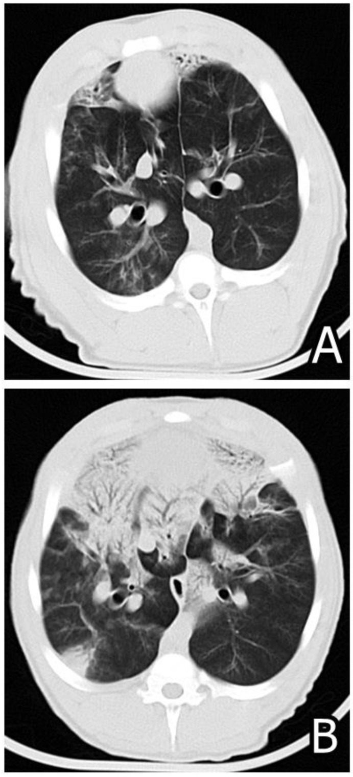
Figure 7.
(A) Computed tomography (CT) photo of lung damages in M. hyopneumoniae experimentally compromised pig in a definite sectioning plane done on day 58 of the experiment showing small focal damages with patchy ground glass opacification. (B) CT photo of the lung damages in M. hyopneumoniae experimentally compromised pig treated additionally with 20 mg/kg FB1 via the consumed feed conducted in the same sectioning plane on day 58 of the experiment showing severe worsening of pneumonic changes as seen from the enlargement of the same damage [131,132].

