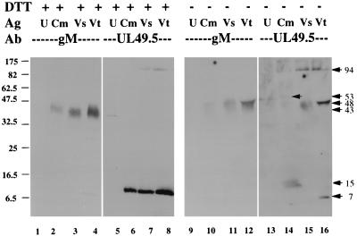
FIG. 8.
Analysis of the gM-UL49.5 complex by Western blotting. Cells were infected at an MOI of 10 and collected 24 h later, and membranes were prepared (Cm). Mock-infected cells were collected at the same time, and membranes were similarly prepared (U). Virions were prepared from cells infected for 60 h at an MOI of 0.5 and either semipurified (Vs) or banded on a potassium tartrate gradient (Vt). Lysates were incubated at 56°C in the presence (lanes 1 to 8) or absence (lanes 9 to 16) of 40 mM DTT for 10 min, analyzed by SDS-PAGE in 10 to 20% gradient gels, and transferred to nitrocellulose paper. The blots were probed with UL49.5T antibody (Ab) first. Bound antibody was detected by ECL (Amersham). The blots were stripped, reprobed with gM antibody, and again detected by ECL. Positions of molecular mass markers on the (left) and calculated sizes of specific bands are indicated on the (right), both in kilodaltons, are indicated. Ag, antigen.

