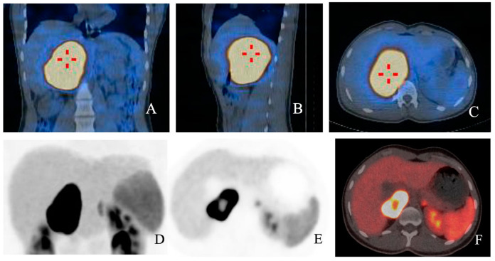
Figure 2.
(A–F): Fusion images with 123I MIBG SPECT/CT in coronal (A), sagittal (B) and axial planes (C) respectively showing high radioligand uptake in a young patient diagnosed with PCC. In (D,E), functional images from the same patient show high uptake of 68Ga-DOTA-SSA, with a central cold area. In fusion image (F), a central spared area of necrosis is present.

