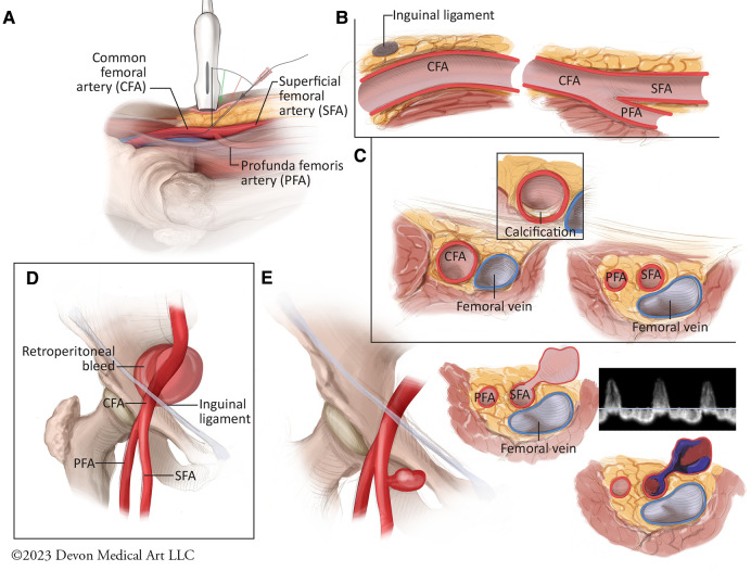
Figure 3.
Best practices for femoral arterial access and potential vascular complications. (A) The optimal technique for ultrasound-guided femoral arterial access requires the use of a 5- to 10-MHz linear ultrasound probe which is held such that the vertical marker is facing upward from the femoral artery to facilitate a longitudinal view of the common femoral artery and its branches. The access needle should then be used at a 45 degree angle to access the common femoral artery. (B) Longitudinal view of the femoral vasculature demonstrates the posterior dive that the common femoral artery takes below the inguinal ligament as the probe is advanced cranially. (C) Rotating the ultrasound proble 90 degrees allows for cross sectional view of the femoral bifurcation, including anterior or posterior vessel wall calcification. (D) Vessel puncture cranial to the inferior epigastric artery is associated with increased risk for retroperitoneal bleeds. (E) Vessel puncture below the common femoral artery bifurcation and involving the superfical femoral artery is associated with increased for pseudoaneurysm formation. Pseudoaneurysms can present clinically as pulsatile hematomas, painful ecchmyoses or as frank bleeding. They typically consist of 1 or 2 layers of the vessel wall, and are therefore associated with increased risk for rupture, embolization, and limb ischemia. They typically demonstrate “to-and-fro” patterns of flow with pulsed Doppler assessment.

