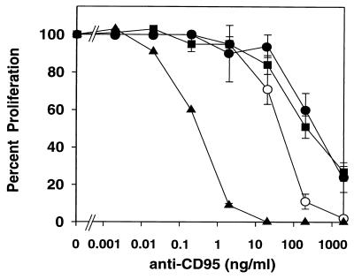
FIG. 6.
Quantification of the effects of CD95 ligation. HVS-transformed T-cell clones ES-BP8T and SS-BP8T, their native parental T-cell clones, and Jurkat cells were compared. The native T-cell clones have been activated with irradiated HLA-compatible PBMC and myelin basic protein. One portion of the native T-cell clones received MAb CH-11 to CD95 1 day after restimulation, and another portion of the native T cells was treated with this MAb 6 days after activation. Spontaneous autocrine proliferation was assessed for the HVS-transformed T cells and the Jurkat cells. About 24 h after addition of the MAbs, the cultures were labeled with [3H]thymidine. The results obtained with the two native T-cell clones treated 1 day after activation (solid circles) or 6 days after activation (open circles), the two HVS-transformed T-cell clones (squares), and Jurkat cells (triangles) were combined. The proliferation in the absence of anti-CD95 was set as 100%, and the inhibition (± standard error of the mean [SEM]) was calculated. Three to six independent experiments were performed per cell line.

