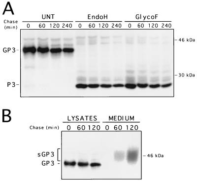
FIG. 3.
Processing and location of PRRSV GP3 protein individually expressed in 293 cells with the recombinant AdCMV5/ORF3 virus. (A) Adherent 293 cells at 80% confluency (107) were infected with AdCMV5/ORF3 at an MOI of 20 to 30 TCID50/cell. At 24 h p.i., cells were pulse-labelled with [35]methionine for 30 min (time zero), chased for the indicated times, and processed for radioimmunoprecipitation with α3 antiserum. Aliquots of immunoprecipitates were treated with Endo H or Glyco F or were left untreated (UNT) and were then analyzed by SDS–12% PAGE. The data indicated that Ad-expressed GP3 was much more stable than PRRSV-expressed GP3. (B) Cells infected with the recombinant AdCMV5/ORF3 virus were pulse-labelled for 30 min and chased for the indicated times as described for panel A. Expression of GP3 was monitored by radioimmunoprecipitation in either the cell lysates or the culture medium. Numbers on the right indicate the positions and sizes of 14C-radiolabelled marker proteins.

