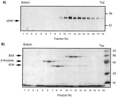
FIG. 4.
Sucrose gradient analysis of e55K. e55K, BSA, β-amylase, and ADH were centrifuged together through a 5 to 20% sucrose gradient. Fractions were collected, trichloroacetic acid precipitated, and analyzed by SDS-PAGE, followed by Western blotting with anti-55K antibody 2A6 (A) and Coomassie blue staining (B). The top and bottom of the gradient are indicated. Molecular mass markers are indicated on the right in kilodaltons. Protein molecular mass standards from the bottom of the gradient are β-amylase (8.9S; 200 kDa; tetramer), ADH (7.4S; 150 kDa; tetramer), and BSA (4.3S; 68 kDa; monomer).

