Figure 5.
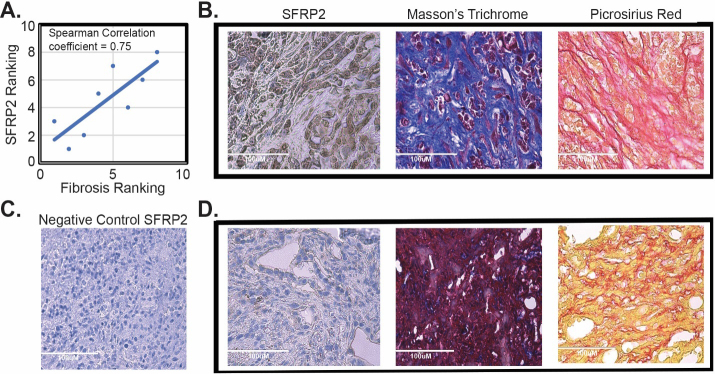
SFRP2 expression is associated with fibrosis in vivo. (A) Correlation of SFRP2 staining and degree of fibrosis in tumors from KPC mice ( 8). (B) Representative images of staining from a KPC mouse that had high SFRP2 staining and high levels of fibrosis identified by Masson’s trichome blue staining and picrosirius red staining. (C) Negative control for SFRP2 immunohistochemistry staining. (D) Representative images of staining from a KPC mouse that had low SFRP2 staining and low levels of fibrosis identified by Masson’s trichome purple staining and picrosirius yellow staining. All slides at 40X HPF: High Power Field. Scale bar: 100 m.

