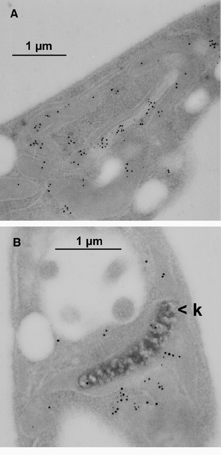
Figure 7.

Double immune electron microscopy. Leishmania cells that express CPN10::GFP chimera were embedded and microsections were prepared. After staining with anti-GFP Mab and anti-mouse immunogold (5 nm), sections were treated with anti-CPN60 antibodies, rabbit anti-chicken IgG, and Protein A gold (10 nm). Thus, 10 nm gold particles represent CPN60 and 5 nm gold particles represent CPN10::GFP. k = kinetoplast.
