Figure 1.
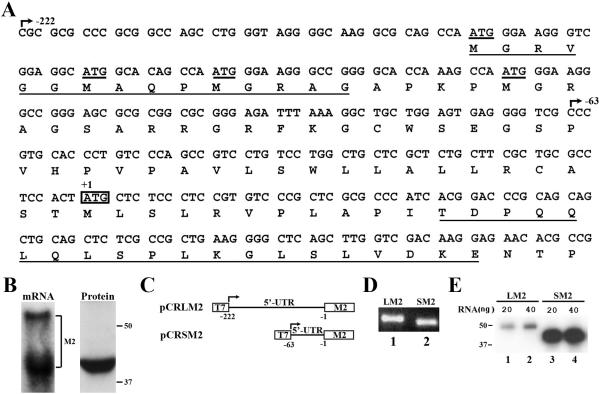
In vitro translation of M2 mRNA with different 5′-UTRs. (A) The DNA sequence encoding the 5′-UTR of M2 mRNA. The upstream AUG codons were underlined and the physiological start codon was boxed. The transcription start sites for mRNAs with both long and short 5′-UTRs were marked with arrows. The protein sequence encoded by the 5′-UTR is also shown with the underlined sequence used for antibody production. (B) Expression of endogenous M2 mRNA and protein in HeLa cells. Northern and western blot analyses were conducted to determine the mRNA and protein species, respectively. (C) Schematic representation of expression constructs with two different 5′-UTRs of M2 cDNA. T7 promoter and the M2 ORF are boxed. Arrows indicate the transcription start site. (D) RNA transcripts. The RNAs were generated from linearized M2 expression constructs by in vitro transcription using T7 RNA polymerase. (E) M2 protein generated by in vitro translation in RRL using the in vitro RNA transcripts as template shown in (D). The products were labeled with [35S]methionine and detected as described in Materials and Methods. LM2, M2 with long 5′-UTR; SM2, M2 with short 5′-UTR.

