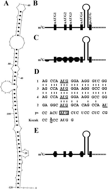
Figure 9.
Model for translational regulation of M2 mRNA with the long 5′-UTR. (A) Secondary structure of 5′-UTR sequence between −120 and the physiological start codon AUG. The secondary structure was predicted using an online program (http://www.bioinfo.rpi.edu/~zukerm/rna/). (B) Schematic representation of the secondary structure (hairpin) relative to the upstream and the physiological start AUGs (filled boxes) in the long 5′-UTR of M2 mRNA. (C) Model of inhibition of translation initiation at the physiological start codon. The secondary structure severs as a barrier for the moving of ribosomes during elongation with initiation at one of the upstream AUGs. (D) Alignment of the AUG triplets with their flanking sequences. The consensus Kozak sequence is also shown. (E) Model of protein-bound upstream AUG triplets. An hypothetical protein (filled circle) binds to an upstream AUG triplet with conserved flanking sequences, which blocks the movement of ribosomes during the elongation of translation initiated at an upstream AUG.

