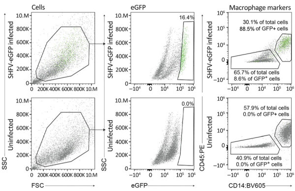
Figure 3.

Flow cytometry of SHFV-eGFP–infected (top plots) and uninfected (bottom plots) splenocytes in study of isolation of diverse simarteriviruses causing hemorrhagic disease. Green dots indicate cells infected with SHFV-eGFP. BV605, brilliant violet 605 dye; eGFP, enhanced GFP; FSC, forward scatter; GFP, green fluorescent protein; PE, phycoerythrin; SHFV, simian hemorrhagic fever virus; SSC, side scatter.
