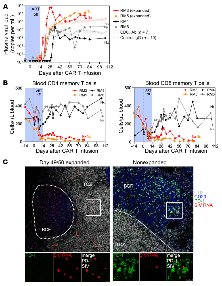
Figure 5. Anti–PD-1 CAR T cell–mediated depletion of TFH cells and PD-1+ T cells does not prevent viral recrudescence after removal of ART.
Plasma viral load in the 4 animals during anti–PD-1 CAR T cell treatment and after removal of ART. Historical controls are shown for the indicated number of RMs as mean (+SEM) (A). Absolute count of CD4+ memory and CD8+ memory T cells in blood (B). Combined immunofluorescence and RNA FISH staining on LN tissue section for CD3 (gray), CD20 (blue), PD-1 (green), and SIV RNA (red). The white line demarcates the border between the T cell zone (TCZ) and the B cell follicle (BCF) (C). Original magnification, ×40.

