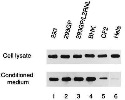
FIG. 1.
Intracellular and extracellular VSV-G in cell lines transfected with pCMV-G. Cells were grown in six-well plates and transfected with plasmid pCMV-G. VSV-G samples in the conditioned medium and cell lysate were examined by established Western blotting methods, using P5D4 monoclonal antibody to visualize VSV-G protein as described in Materials and Methods.

