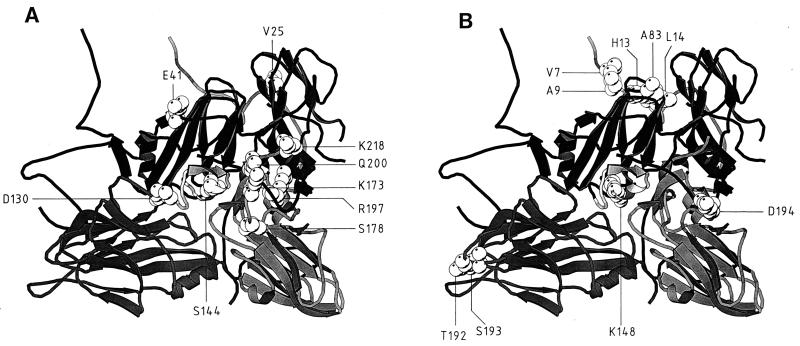
FIG. 5.
Locations of the amino acid substitutions found in FMDV MARLS (A) and FMDV R100 (B) on a ribbon protein diagram of a crystallographic protomer of C-S8c1 (33). The capsid proteins VP1, VP2, and VP3 are represented as dark, medium, and light grey, respectively. VP1 from a neighboring protomer is shown at the upper right. The position of the G-H loop of VP1 in C-S8c1 corresponding to that found in the complex with MAb SD6 is shown in light grey at the center of each structure (27). The substituted residues are depicted in van der Waals spheres. The amino acids indicated are those listed in Table 5 for MARLS and R100. The minimal side chain-side chain distances measured between critical amino acid residues of MARLS and were 3.6 Å between VP2 D-130 (Cδ2) and VP1 A-145 (N), 4.5 Å between VP3 K-173 (Nζ) and VP1 Q-200 (Oɛ1), 4.7 Å between VP3 K-173 (Cδ) and VP1 R-141 (Nɛ), 5.1 Å between VP1 Q-200 (Cα) and VP1 R-141 (Cζ), 12.7 Å between VP1 R-197 (Cα) and VP1 R-141 (Nη2), and 15.0 Å between VP3 S-178 (Cβ) and VP1 P-156 (Cγ). The corresponding Cα-Cα distances were 6.8, 5.6, 9.5, 9.7, 17.9, and 16.5 Å, respectively. A minimal side chain distance of 13.3 Å was measured between R100 VP1 D-194 (Oδ2) and VP1 R-141 (Nη2). The corresponding Cα-Cα distance was 21.1 Å. The origin of the viruses and the procedures used to locate amino acids on the capsid structure are described in Materials and Methods.

