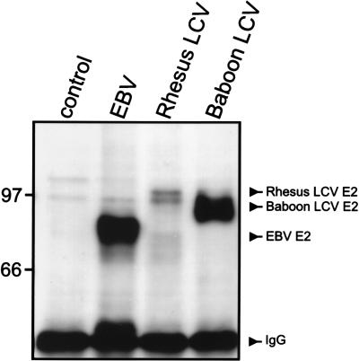
FIG. 4.
EBNA-2 expression in EBV-negative B-lymphoma cells, BL41, after acute infection with control medium, EBV, rhesus monkey LCV, and baboon LCV. The EBV (88 kDa), rhesus monkey LCV (100 kda), and baboon LCV (94 kda) EBNA-2 proteins were immunoprecipitated from cells 2 days after infection and detected by Western blotting with the monoclonal antibody PE2, which recognizes a conserved EBNA-2 epitope. E2, EBNA-2; IgG, immunoglobulin G. Molecular mass markers (kilodaltons) are at left.

