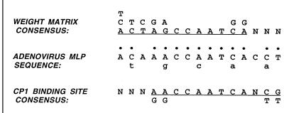
FIG. 2.
Sequence of the CAAT box of the MLP compared with the consensus derived either from a weight matrix analysis (5) of eukaryotic promoters or a consensus (7) from promoters shown experimentally to bind CP1. The single bases shown above the weight matrix consensus correspond to those found in at least 20% of the promoters analyzed. The single bases shown below the CP1 binding consensus are alternative residues found in more than one promoter of high affinity. The dots indicate those residues in the adenovirus MLP that correspond to one or both consensus sequences. Bases shown in lowercase below the MLP sequence are the mutations in CAAT5.

