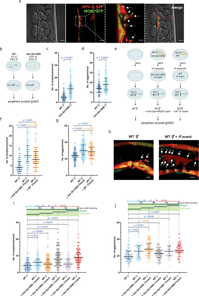
Fig. 1. Production of C. elegans muscle exophers is regulated by male pheromones.
a C. elegans muscle exophers featuring mitochondria, shown with red (RPN-5::wrmScarlet proteasome subunit) and green (mitochondrial outer membrane::GFP) fluorescence. Arrows point to exophers, asterisks mark mitochondria-containing exophers (consistent in at least 25 animals from three replicates). b Experimental setup schematic: him-5(e1490) mutant males were co-cultured from the L1 stage until the L4 stage and then transferred to a male-free plate until AD2 for exopher count assessment. Created with BioRender.com. c Increased percentage of males in the population (via him-5(e1490) mutation) leads to a higher exophergenesis level in hermaphrodites. n = 79 and 83 worms, N = 3 independent experiments. d Increased percentage of males in the population (via him-5(e1490) mutation) causes embryo retention in hermaphrodites. n = 79 and 85 worms, N = 3 independent experiments. e Experimental setup schematic: 50 him-5/wild-type males cultured on a plate for 48 h, then removed. Subsequently, 10 hermaphrodites were transferred to these plates and grown until AD2 for exopher number assessment. Created with BioRender.com. f Growing wild-type hermaphrodites on him-5(e1490) or wild-type male-conditioned plates increases exophergenesis levels. n = 106, 98, and 89 (for respective columns), N = 3 independent experiments. g Exposing wild-type hermaphrodites to him-5(e1490) or wild-type male secretome causes embryo retention in the uterus. n = 105, 98, and 60 (for respective columns), N = 3 independent experiments. h C. elegans hermaphrodites increase exopher production upon male pheromone exposure, shown with red (RPN-5::wrmScarlet proteasome subunit) and green (mitochondrial outer membrane::GFP) fluorescence. Arrows point to exophers, asterisks mark mitochondria-containing exophers (consistent in at least 25 animals from three replicates). i–j Exposure to male pheromones for 24 h triggers exophergenesis in hermaphrodites, regardless of the life stage at which the exposure occurs. Higher embryo retention in the uterus coincided with an increase in exopher production. i n = 90, 87, 85, 88, 89, and 90 worms (for respective columns), N = 3 independent experiments. j n = 50, 50, 48, 50, 50, and 50 (for respective columns), N = 2 independent experiments. Data information: Scale bars are (a) 20 μm and 5 μm (zoom in), h 10 μm. @AD2 means “at adulthood day 2”. Data are presented as median with interquartile range; not significant p values (p > 0.05) are in orange color, significant p values (p < 0.05) are in blue color (c–d) two-tailed Mann–Whitney test, (f–g, i, j) Kruskal-Wallis test with Dunn’s multiple comparisons test. Source data are provided as a Source Data file.

