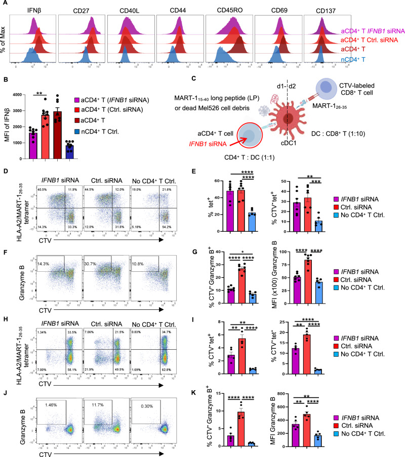
Fig. 4.
IFN-I produced by activated CD4+ T cells is important for the cDC1-mediated CTL response to cell-associated tumor antigens. A tumor antigen-specific CTL priming system [6] was used to investigate the impact of IFN-I produced by activated CD4+ T cells on the cross-presentation and cross-priming abilities of cDC1s. IFNβ expression was downregulated in CD4+ T cells by siRNA. A Flow cytometry histograms depicting intracellular IFNβ expression and the surface expression of the indicated markers identifying effector T cells under the indicated conditions. B MFI values for intracellular IFNβ expression under the indicated conditions. C Schematic illustration of the tumor antigen-specific CTL priming system. D CD8+ T-cell proliferation induced by MART-115–40 long peptide (LP) based on CTV dilution. E Percentages of MART-126–35/HLA-A2-specific (tetpostive) cells among CD8+ T cells or CTVnegative CD8+ T cells in the MART-115-40 LP setting. F CTL response to MART-115–40 LP based on intracellular Granzyme B staining. G Percentages of Granzyme B+ cells among CTVnegative CD8+ T cells and MFI values of Granzyme B in the MART-115-40 LP setting. H CD8+ T-cell proliferation in response to dead Mel526 cell debris based on CTV dilution. I Percentages of MART-126-35/HLA-A2-specific (tetpositive) cells and tetnegative cells among CTVnegative CD8+ T cells in the dead Mel526 cell debris setting. J CTL response to dead Mel526 cell debris based on intracellular Granzyme B staining. K Percentages of Granzyme B+ cells among CTVnegative CD8+ T cells and MFI values of Granzyme B in the dead Mel526 cell debris setting. The data were pooled from eight (n = 8 in B), seven (n = 7 in E, G) or five (n = 5 in I, K) independent experiments. P < 0.05*, P < 0.01**, P < 0.001***, P < 0.0001**** (one-way ANOVA). The data are shown as the means ± SEMs

