Fig. 5.
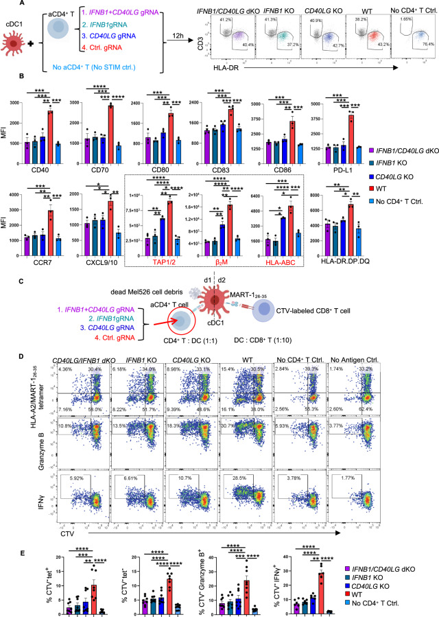
CD4+ T-cell help provided via IFNβ rather than via CD40 signaling promotes MHC-I antigen cross-presentation in cDC1s. Purified naive CD4+ T cells were transfected with Cas9/ctrl. gRNA, Cas9/IFNB1 gRNA, Cas9/CD40LG gRNA or the Cas9/IFNB1+CD40LG gRNA ribonucleoprotein (RNP) complex were subsequently stimulated with anti-CD3/CD28 antibodies. A Schematic illustration of the cDC1-CD4+ T-cell coculture system and the gating strategies for flow cytometric analysis of the cDC1 response. B MFI values for the expression of the indicated cDC1 “help” signature markers under the indicated conditions. The black box highlights the molecules involved in MHC-I antigen presentation. C–E The tumor antigen-specific CTL priming system [6] was used to investigate the impact of IFN-I and CD40L produced by CD4+ T cells on cDC1-mediated CTL priming. Dead Mel526 cell debris was used as an antigen source. C Schematic illustration of the experimental procedures. (D) CD8+ T-cell proliferation based on CTV dilution and the CTL response based on intracellular Granzyme B and IFNγ staining. E Percentages of MART-126-35/HLA-A2-specific (tet+) cells, tet(-) cells, and Granzyme B+ or IFNγ+ cells among CTV(-)CD8+ T cells. The data were pooled from three (n = 3 in B) or 8 (n = 8 in E) independent experiments. P < 0.05*, P < 0.01**, P < 0.001*** (one-way ANOVA). The data are shown as the means ± SEMs

