Figure 5.
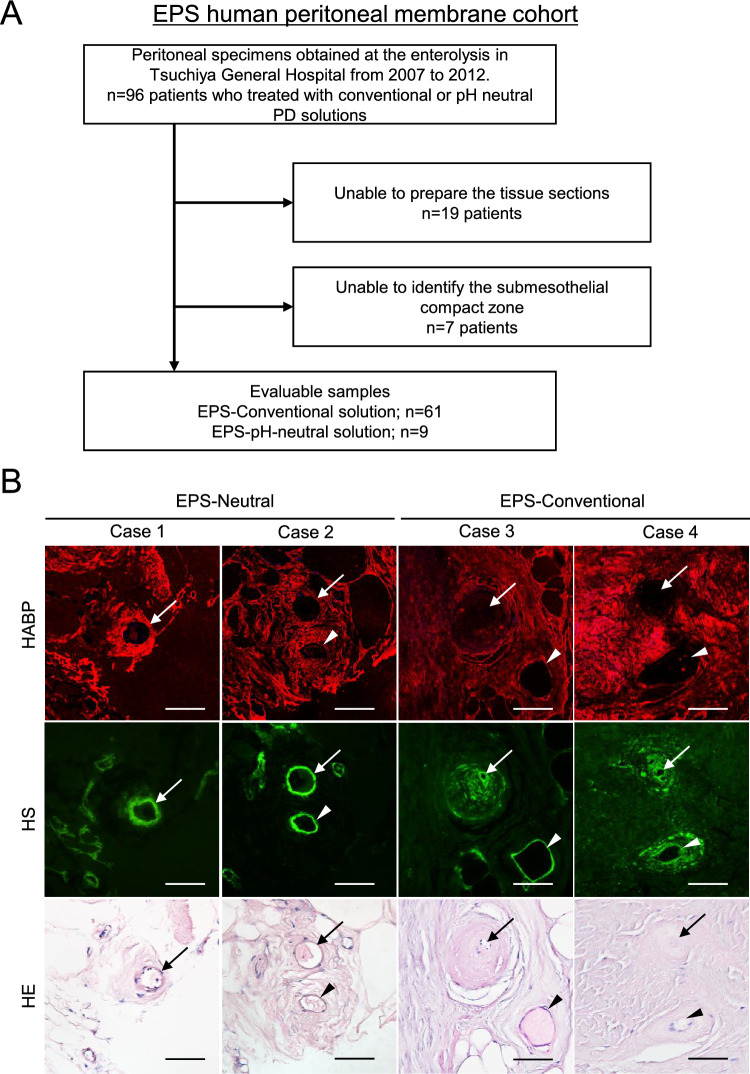
Expression of hyaluronan in EPS human peritoneal membrane. (A)Flow diagram of the study population. (EPS human peritoneal membrane cohort). (B) HABP, HS, and HE staining. (C) Differences in L/V ratio, HABP and HS positivity, CD31-positive vessels, and CD68-positive cells. (D) Expression on CD31-positive vessels and CD68-positive macrophages. Two representative cases are shown in each group. HE staining was conducted after HABP staining on the same sections. Arrows and arrowheads indicate the same vessels in each case. Expression of hyaluronan assessed by HABP positivity was decreased in association with lower L/V ratio in EPS cases treated with conventional solution. HS was more preserved than hyaluronan, as assessed by HABP staining. A correlation between L/V ratio and HABP positivity was observed (r2 = 0.474, p < 0.0001). The number of CD31-positive vessels was higher in the EPS-neutral group. Control: (green circle); EPS-Conventional: EPS cases treated with conventional solution during PD (red circle); EPS-Neutral: EPS cases treated with low-GDP, pH-neutral solution during PD (blue circle); L/V ratio: luminal diameter to vessel diameter; HABP hyaluronan-binding protein, LEL Lycopersicon esculentum lectin, HS heparan sulfate, n.s. not significant. Scale bars = 100 μm.


