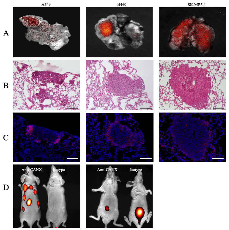
Fig. 3.
In vivo and ex vivo imaging of a tumor xenograft model. (A) Ex vivo imaging of lungs in a tumor xenograft mouse model. (B) Lung tissues stained with H&E for ex vivo imaging. (C) Serial sections stained with DAPI and observed under a fluorescence microscope (× 100; scale bar, 100 μm). (D) In vivo imaging of a subcutaneous tumor xenografted model. Imaging results were obtained at 3 hours after intravenous injection of labeled antibodies (n = 6).

