
Figure 21.
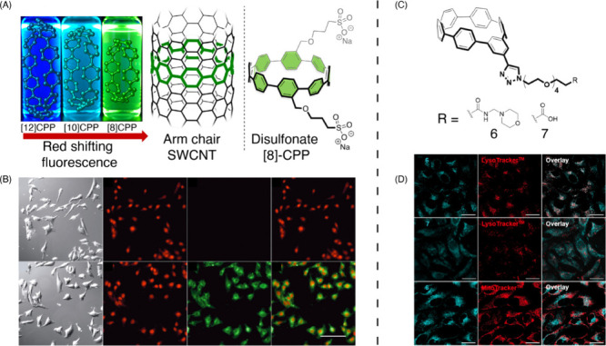
Live cell imaging using carbon nanohoops. (A) CPPs can be conceptualized as the smallest macrocyclic slices of an armchair nanotube. Notice the counter-intuitive red shifting of fluorescence as ring size decreases. Right: structure of cell permeable disulfonate [8]CPP, used for live cell imaging depicted in panel B. (B) Bright-field, nuclear (NucRed, red) and cytoplasmic (disulfonate [8]CPP, green) images of HeLa cells, and overlay between red and green channels. Top row: imaged in the absence of disulfonate [8]CPP. Reproduced from ref (1011). Copyright 2018 American Chemical Society. (C) Structure of meta[6]CPP with PEG chains to enhance aqueous solubility, capped with subcellular targeting ligands (R). (D) Top row: Lysosome-targeting motif enables localization of meta[6]CPP punctate signal to lysosome (good overlap with LysoTracker). Middle row: Nanohoop without lysosome-targeting motif exhibits diffuse labeling and poor overlap with LysoTracker. Bottom row: lysosome-targetted nanohoops show poor overlap with MitoTracker, a mitochondrial marker. Reproduced from ref (1013). Copyright 2021 American Chemical Society.