Figure 1:

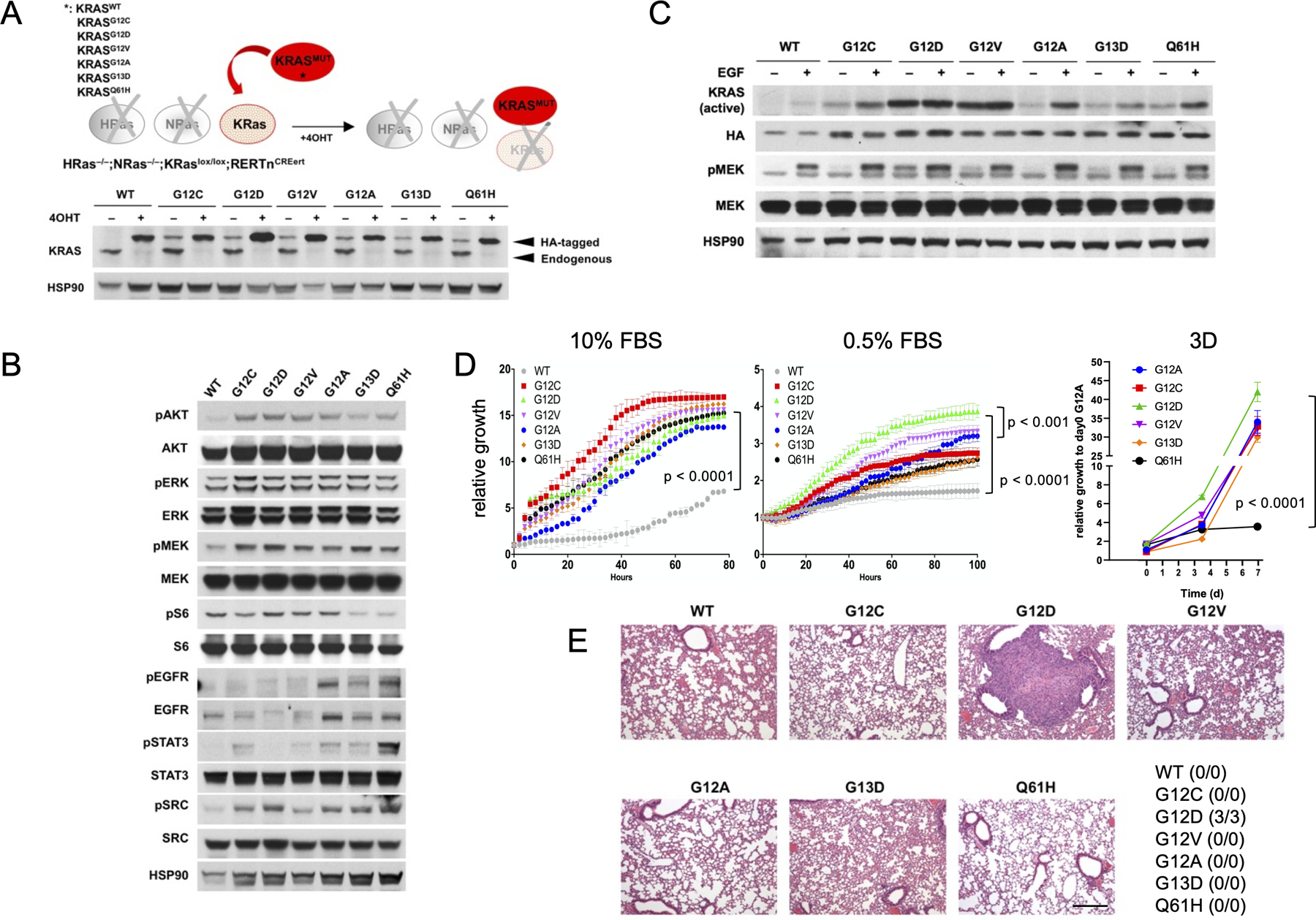
(A) schematic representation of the KRaslox KRASMUT system (top panel). HRas–/–; NRas–/–; KRaslox/lox mouse embryonic fibroblasts were infected with 5-MOI of retroviruses encoding for different human HA-tagged KRAS mutants, selected with puromycin (1μg/ml), treated with 4OHT (600nM) for at least 10 days and probed by Western Blot with the indicated antibodies (bottom panel). (B) KRaslox KRASMUT cells were treated with 4OHT for 10 days and probed by Western Blot with the indicated antibodies. Results are representative of one of three similar experiments. (C) KRas-GTP levels and activation of downstream pMEK signaling in KRaslox KRASMUT cells in 0.1% FBS medium upon stimulation with EGF (50 ng/mL). Results are representative of one of three similar experiments. (D) Growth rates of KRaslox KRASMUT cells in 10% or 0.5% fetal bovine serum (FBS) medium in 2D conditions as assessed by IncuCyte measurements (left panels, p < 0.0001 by unpaired Student’s t test). Results are representative of one of three similar experiments. Growth of KRaslox KRASMUT organoids was monitored by CTG assay. ANOVA analysis followed by Tukey’s multiple comparisons post-test was used for statistical analysis (p < 0.0001). (E) KRaslox KRASMUT cells (1×10⁶) were injected into the tail vein of nude mice (n=3 per isoform). Animals were sacrificed after one month and lung colonization checked by serial sections stained with hematoxylin and eosin. The number of animals with evidence of lung colonization is indicated in the table. Scale bar: 100μM.
