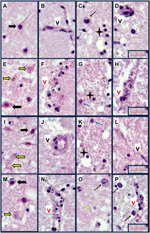
Fig. 2.
Cellular Neuropathology of FLSCs treated with 2,4-D, 2,4,5-T, or D + T. Formalin-fixed paraffin-embedded, H&E-stained histological sections of FLSCs treated with A–D) Vehicle, E–H) 250 μg/ml of 2,4-D, I-L) 250 μg/ml of 2,4,5-T, or M–P) D + T (both agents) for 24 h. Cortex (Panels A, B, E, F, I, J, M, N) showing relatively intact neurons (black arrows) or neurons with degenerative of apoptotic pathology (yellow arrows), and relatively intact vessels (Black ‘V’) or vascular necrosis (Red ‘V’). White matter (Panels C, D, G, H, K, L, O, P) with normal round oligodendrocytes (black 4-point stars) or oligodendrocyte apoptosis (yellow 4-point star), reactive enlarged astrocyte (black arrows) or injured, eosinophilic astrocyte (blue arrow), intact vessels/vascular endothelial cells (Black ‘V’) or degenerated necrotic vessels (Red ‘V’). Samples were photographed at 600x, then cropped and re-scaled. Final image magnification scale bars are displayed for each row.

