
Figure 2. ApoER2 binds via an IXXF/Y motif to a canonical site on the μ4 subunit of AP-4.
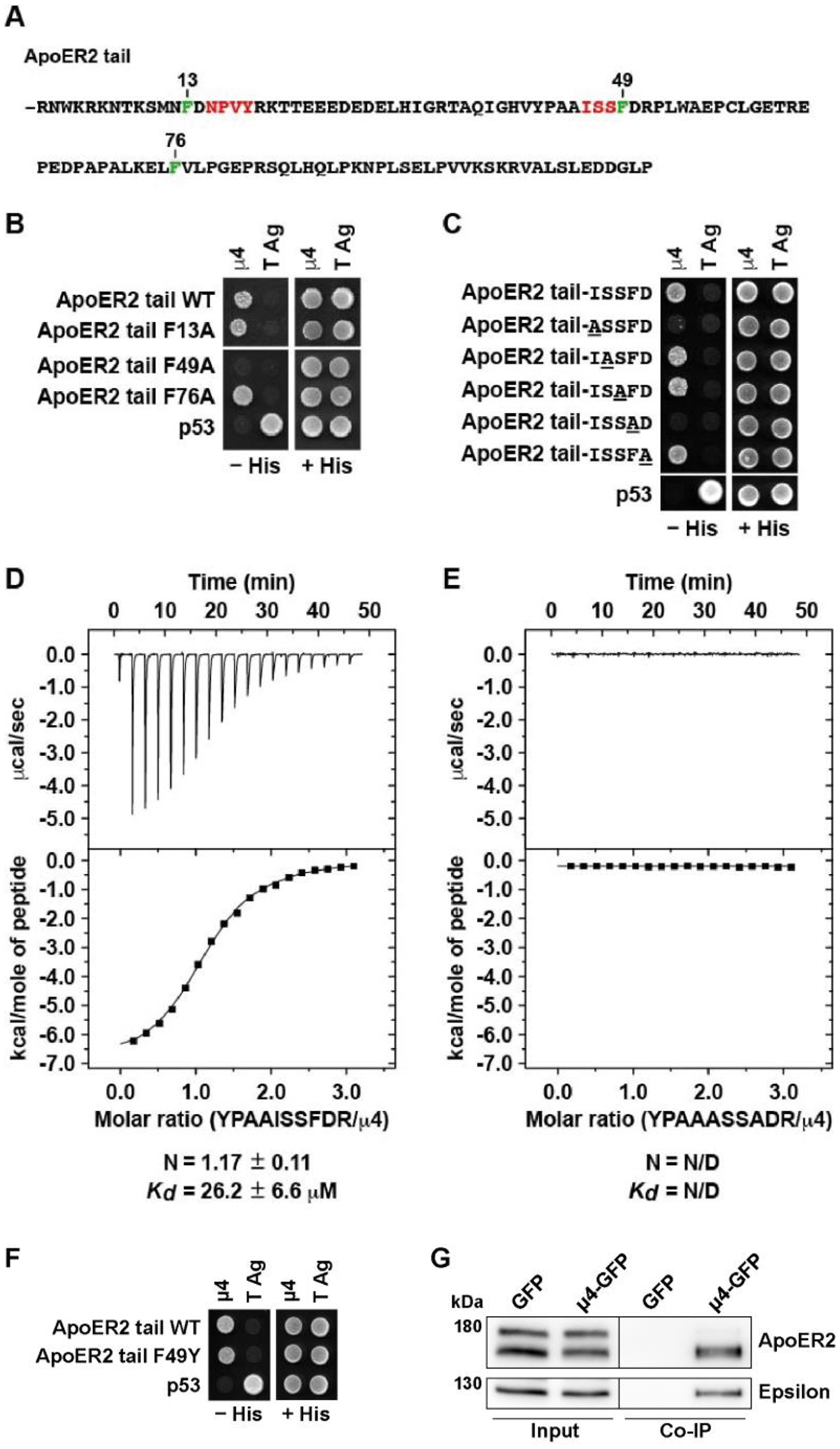
(A) Sequence of the ApoER2 cytosolic tai (residues 849–963), highlighting in green three phenylalanine residues at positions 13, 49 and 76 relatives to R849 as residue 1 of the indicated cytosolic tail sequence. Highlighted in red are an NPXY motif and the sequence ISS adjacent to F49 that is important for interaction to μ4. (B, C) Y2H analysis of critical residues on the ApoER2 cytosolic tail for interaction with μ4. Yeast cells were co-transformed with plasmids encoding Gal4bd fused to wt or the indicated mutants of the ApoER2 cytosolic tail, and Gal4ad fused to the wt μ4 subunit of the AP-4 complex. Mouse p53 fused to Gal4bd and SV40 large T antigen (T Ag) fused to Gal4ad were used as controls. Co-transformed cells were spotted onto His-deficient (-His) or His-containing (+His) plates and incubated at 30 °C. (D-E) Isothermal titration calorimetry of YPAAISSFDR peptide (D) or YPAAASSADR peptide (E) with recombinant μ4 C-terminal domain. The Kd and stoichiometry (N) for the μ4-YPAAISSFDR interaction are expressed as the mean ± SD (n = 3). N/D: not determined. (F) Y2H analysis of the effect of substituting tyrosine for F49 in the cytosolic tail of human ApoER2 on interaction with μ4. The analysis was performed as described for panels B and C. (G) Wild-type HeLa cells were co-transfected with plasmids encoding FLAG-tagged mouse ApoER2 and μ4-GFP or GFP as control. Twenty-four h later, cells were lysed, and cell extracts were immunoprecipitated using GFP-trap. The presence of the receptor was detected by immunoblotting. Notice that the receptor was present in both cell extracts (8% of input) but was immunoprecipitated only from cells expressing μ4-GFP.