
Figure 10. Images demonstrating examples of stage 5 retinopathy of prematurity.
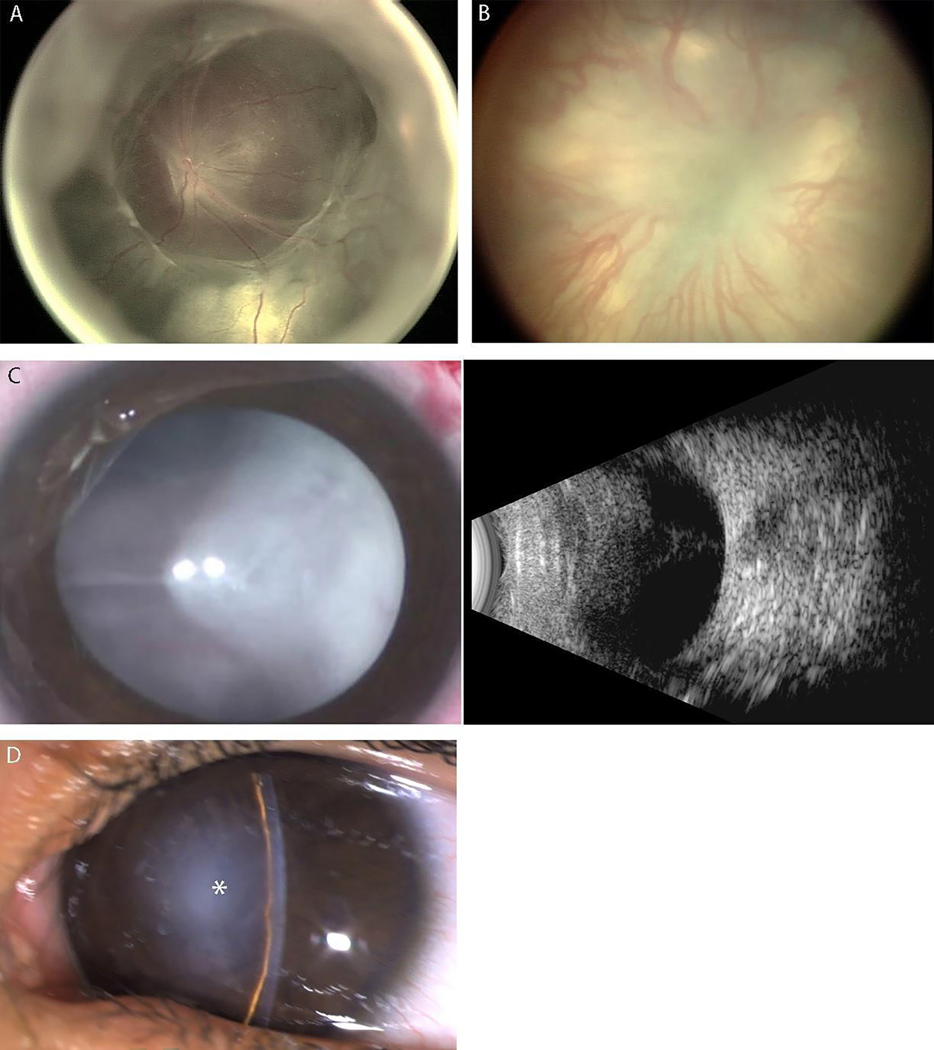
(A) Wide-angle fundus photograph showing stage 5A, characterized by a total retinal detachment with visible optic disc. Note open-funnel configuration. (B) Wide-angle fundus photograph showing Stage 5B, with no view of optic disc because of fibrovascular tissue. (C) External photograph of the normal anterior segment in stage 5B (left side), with no view of optic disc or retina secondary to retrolental fibrovascular tissue. B-scan ultrasonography (right side) reveals total retinal detachment with a posteriorly closed funnel configuration. (D) External photograph showing anterior segment characteristic of Stage 5C with anterior lens displacement, marked anterior chamber shallowing, central irido-capsule-endothelial adhesion, and central corneal opacification (asterisk) that prevent view of closed-funnel retinal detachment.