
Figure 11. Wide-angle fundus photographs demonstrating examples of ROP regression.
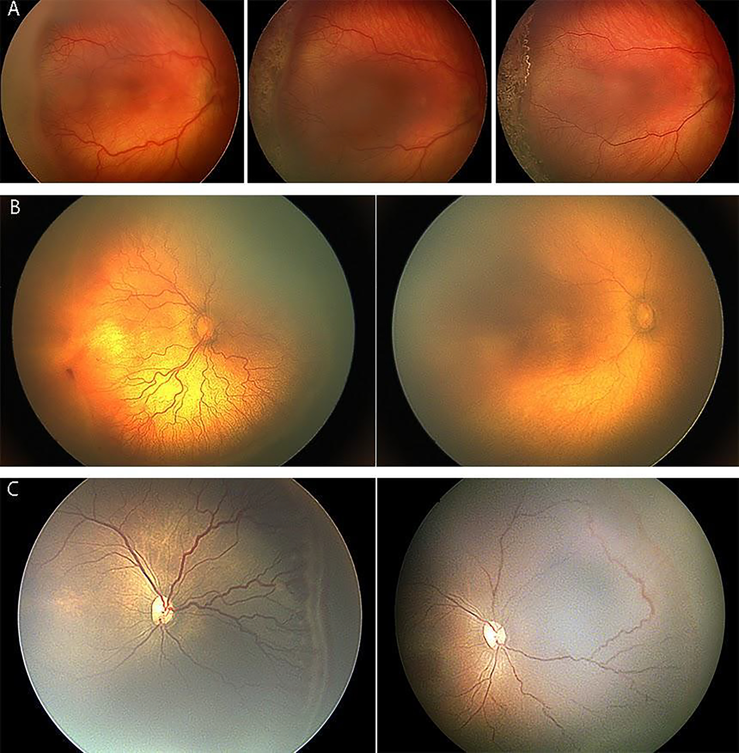
(A) Regression after laser treatment. Image on left is pre-treatment, showing stage 3 with plus disease. Image in middle is 1 week post-treatment, showing that stage 3 is thinner and whiter. Image on right is 1 month post-treatment, showing disease regression. (B) Regression of plus disease after anti-VEGF injection for aggressive ROP (A-ROP). Image on left is pre-treatment, showing plus disease and flat neovascularization (stage 3). Image on right is 2 weeks post-treatment, showing improvement in plus disease with no visible ROP lesion. (C) Regression after anti-VEGF injection. Image on left is pre-treatment. Image on right is 4 weeks post-treatment, showing absence of stage 3 and improvement in plus disease, with vascularization into peripheral avascular retina. Note the circumferential anastomosis in the area of original stage 3, along with reactivated stage 1 more anteriorly.