
Figure 5. Wide-angle fundus photographs demonstrating examples of acute retinopathy of prematurity (ROP) stages 1–3.
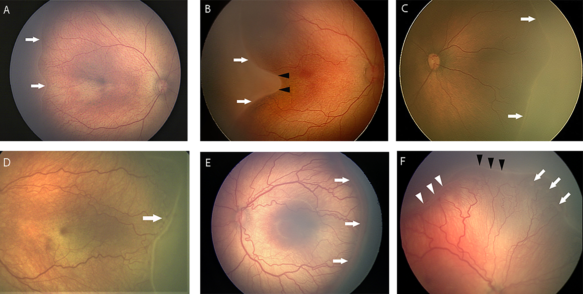
(A) Stage 1 demarcation line at border between vascular and avascular retina (white arrows). (B) Stage 1 demarcation line (white arrows) and associated notch (black arrowheads) between vascular arcades which would be considered zone I secondary to notch. Note pre-plus disease with mild retinal vascular tortuosity and dilation. (C) Stage 2 ridge, which is raised (white arrows) and thicker than stage 1. (D) Stage 2 ridge. Note “popcorn” lesions posterior to ridge (arrows) and pre-plus disease with mild vascular tortuosity and dilation. (E) Stage 3 disease with extraretinal neovascularization (white arrows). Note plus disease with vascular tortuosity and dilation. (F) Eye with both stage 2 (black arrowheads) and stage 3 (white arrowheads), and associated “popcorn” (white arrows). Note plus disease with vascular tortuosity and dilation.