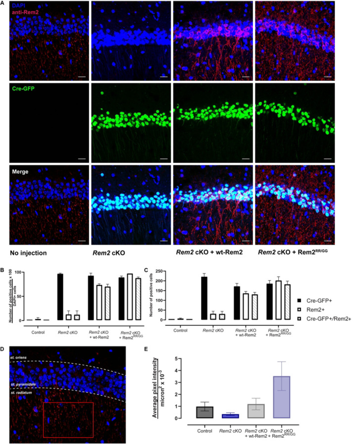
Figure 2.
Viral-mediated conditional knockout of Rem2 and rescue with either wt-Rem2 or Rem2RR/GG in hippocampus from P28 mice. A) Representative images from coronal sections of the dorsal CA1 from uninjected and virus-injected mice. Top, DAPI and anti-Rem2; middle, Cre-GFP; bottom, merged image. DAPI = blue, Cre-GFP = green, anti-Rem2 = red. B) Quantification of the percentage of neurons expressing indicated markers divided by DAPI-positive neurons for each experimental condition. C) Total number of neurons expressing indicated markers for each condition. D) Schematic for quantifying anti-Rem2 staining in the dendritic arbor of pyramidal neurons, from an uninjected control animal. A region of interest was drawn (red rectangle) in the striatum radiatum within the CA1 region of the hippocampus, the average pixel intensity in the channel for anti-Rem2 staining was measured. E) Average pixel intensity per square microns for each condition. Error bars = SEM. 350–1000 cells per condition from 2–3 slices per biological replicate in 2 biological replicates. Scale bar = 20 µm. st. = striatum.

