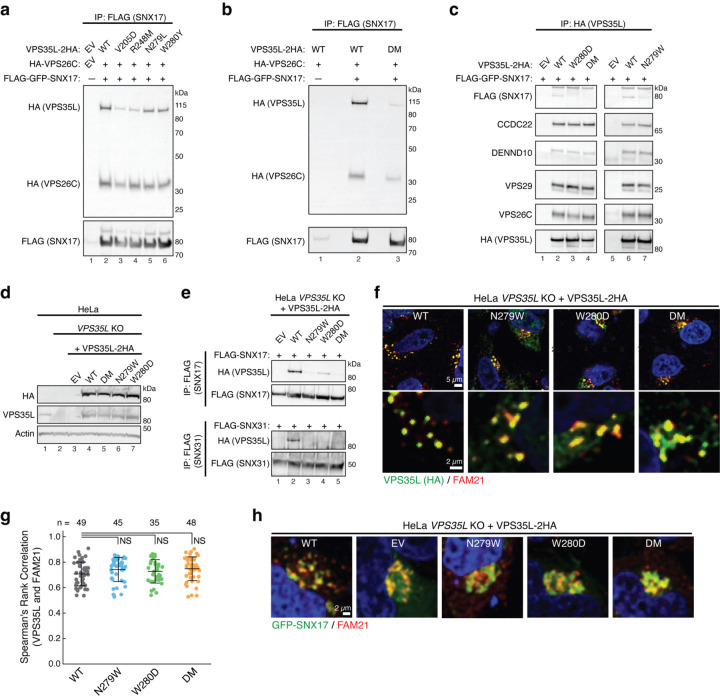
Fig. 3. Disrupting the SBP impairs SNX17 and SNX31 binding in cells.
a-b. Immunoprecipitation of SNX17 (FLAG) followed by immunoblotting for VPS35L and VPS26C (HA) in HEK293T cells transfected with the indicated expression vectors. EV, empty vector; DM, double mutant. c. Immunoprecipitation of VPS35L (HA) followed by immunoblotting for SNX17 (FLAG) and indicated protein components of the CCC and Retriever complexes in HEK293T cells transfected with indicated SNX17 and VPS35L variants. d. Immunoblotting analysis for endogenous and stably expressed VPS35L in the indicated HeLa cell lines derived from a VPS35L knockout (KO) rescued with the indicated variants of VPS35L or an empty vector (EV) control. The parental HeLa cell line used to derive the VPS35L knockout line is included for comparison. e. Immunoprecipitation of SNX17 (top) or SNX31 (bottom) after transfection in the indicated HeLa cell lines, followed by immunoblotting for VPS35L (HA). f-g. Representative confocal images (f) and quantification of colocalization (g) derived from concurrent immunofluorescence staining for VPS35L (HA, green) and the endosomal marker FAM21 (red) in HeLa cells shown in (e). In (g), each dot represents an individual cell, with number of cells in each group indicated above the graph. Mean and standard deviation are shown. One-way ANOVA with Dunnett’s correction was used. NS, not significant. h. Representative confocal images showing concurrent immunofluorescence staining for GFP-SNX17 (green) and the endosomal marker FAM21 (red) in HeLa cells shown in (e) and transfected with GFP-SNX17.

