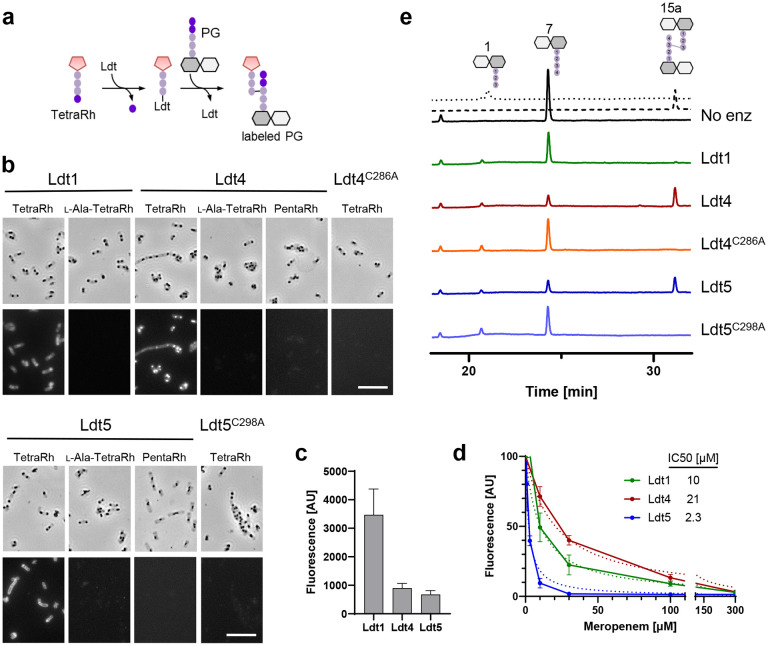
Fig. 3. VanW domains catalyze l,d-transpeptidation in vitro.
(a) Schematic diagram of incorporation of TetraRh into PG sacculi by an Ldt. Pink pentagon, Rhodamine. Colored balls, amino acids. Dark and light gray hexagons, NAM and NAG, respectively. (b) Phase contrast and fluorescence micrographs of immobilized PG sacculi after incubation for 1 h with 5 μM enzyme and 30 μM substrate analog as indicated. TetraRh: LDT-specific substrate analog; l-Ala-TetraRh, negative control; PentaRh, PBP-specific substrate. Size bar, 10 μm. Micrographs are representative of at least two experiments. (c) Quantification of TetraRh incorporation into sacculi graphed as the mean ± s.d. of the fluorescence intensity from 10 sacculi. (d) Inhibition of LDT activity by meropenem graphed as the mean and s.d. of data pooled from four experiments. IC50 is the concentration of meropenem needed to reduce LDT activity by half. (e) HPLC analysis of muropeptides after 1 h incubation of the indicated enzymes with DS-TetraP substrate. Structures above the chromatograms are numbered according to Peltier et al.15. Calibration traces for 1 and 15a are shown by the dotted and dashed lines, respectively. Chromatograms are representative of 3 experiments.

