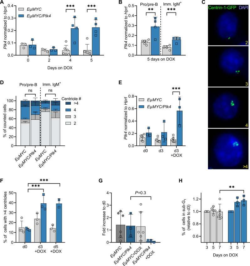
Fig. 2. PLK4-induced centrosome amplification in B cells leads to cell death.
(A) Plk4 transgene expression in whole-blood samples of premalignant EμMYC and EμMYC/Plk4 transgenic mice at the age of 5 weeks, kept on doxycycline for up to five additional days (n = 3 to 6). (B) Plk4 expression in pro/pre-B and immature IgM+D− B cells isolated by cell sorting from spleens of 5-week-old animals kept for 5 days on doxycycline. Expression was normalized to Hprt (EμMYC n = 6; EμMYC/Plk4 n = 5). (C) Example picture of Centrin-1-GFP foci marking centrioles (scale bar, 8 μm) in pro/pre-B and immature IgM+D− B cells after isolation from spleens of 5-week-old EμMYC and EμMYC/Plk4 transgenic mice, quantified in (D). (D) Percentage of EμMYC (n = 3) and EμMYC/Plk4 (n = 4) pro/pre-B and immature IgM+ B cells presenting with 2, 3, 4, or >4 centrioles after 5 days on doxycycline-containing food. (E) Plk4 transgene expression in pro-B cells isolated from EμMYC (n = 6) and EμMYC/Plk4 mice (n = 3), cultured with or without doxycycline (1 μg/ml) for 3 days. Expression was normalized to Hprt. (F) Percentage of EμMYC (n = 2) and EμMYC/Plk4 (n = 2) pro-B cells with >4 centrioles, cultured with or without doxycycline for 3 and 5 days. (G) Fold change of total cell number in pro-B cell cultures on day 7 normalized to day 0 (n = 3 to 6). (H) Percentage of sub-G1 cells in the respective pro-B cell cultures normalized to day 3 on days 5 and 7 (EμMYC n = 5, EμMYC/Plk4 n = 3). Data are shown as means ± SD and were statistically tested by Sidak’s multiple comparisons test. *P < 0.05, **P < 0.01, ***P < 0.005.

