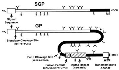
FIG. 2.
Diagrammatic representation of SGP and GP molecules of EBO virus (Zaire species isolated in 1976) showing important structural features. The white N-terminal regions of SGP and GP correspond to identical (shared) sequences, while the black C termini identify sequences unique to GP or SGP molecules. The common signalase cleavage sites for both SGP and GP and the furin cleavage site for GP0 (uncleaved form of GP) (↓) were determined by N-terminal sequencing. Also shown are cysteine residues (S), predicted N-linked glycosylation sites (Y-shaped projections), a predicted fusion peptide, a heptad repeat sequence, and a transmembrane anchor sequence. In EBO viruses, the positions of these structures are conserved and their sequences are very similar or, in the case of N-linked glycosylation sites, are at least concentrated in the central region of GP.

