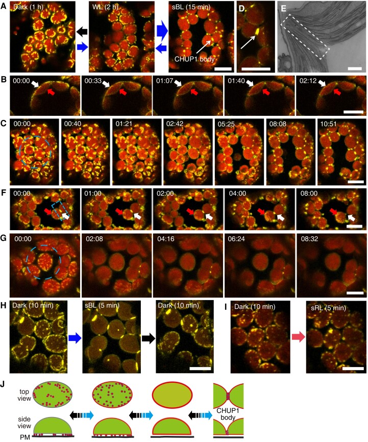
Figure 1.
Reorganization of CHUP1-YFP on the chloroplast envelope in response to blue light. A) Reversible changes in the distribution pattern of CHUP1-YFP under the indicated light conditions. CHUP1-YFP in the palisade cells of C1Y transgenic Arabidopsis plants was observed following incubation in darkness for 1 h (black arrow), weak white light (20 μmol m−2 s−1) for 2 h (thin blue arrow), or strong blue light (sBL; 458-nm laser scan with an output power of 2.8 µW) for 15 min (thick blue arrow). The images were captured at a resolution of 512 × 512 pixels using a 4× digital zoom and are presented with false-color indicating YFP (yellow) and chlorophyll (red) fluorescence. The time (min:s) of image acquisition is shown in the upper left corner of each image. B) Redistribution of CHUP1-YFP from the interface between the plasma membrane and the chloroplast envelope (white arrows) to the entire chloroplast envelope, including the vacuole side (red arrows). The whole cell was irradiated with strong blue light. Time-lapse images were captured at 33-s intervals. The image acquisition and presentation are the same as in A). C) Blue light-dependent reorganization of CHUP1-YFP during a strong light-induced avoidance response followed by dark adaptation for 20 min. The dashed blue circle indicates the area that was irradiated. A time-lapse movie of this response can be seen in Supplementary Movie S1. D) A CHUP1 body appears as 2 lines along the interface between 2 chloroplasts after 5 min of strong blue light irradiation. E) Transmission electron microscopy image of the region of contact (at the center of the rectangle) between 2 chloroplasts. F) Redistribution of a CHUP1 body (white arrows) to small dots (red arrows) at the leading edge of a moving chloroplast. G) Redistribution of CHUP1-YFP on an isolated irradiated chloroplast. The dotted CHUP1-YFP signal rapidly disappeared but no CHUP1 body formed. A time-lapse movie of this response can be seen in Supplementary Movie S2. The area irradiated with strong blue light is indicated as a dashed blue circle (20 µm in diameter) in C and G), and a dashed blue rectangle (10 μm × 5 μm) in F) superimposed on the first image (00:00) of each series. Strong blue light was provided by 458-nm laser scans (an output power of 2.8 μW). Time-lapse images were collected at 40-, 33-, 60-, and 30-s intervals in C, F, and G), respectively. The image acquisition and presentation are the same as in A). H and I) Blue light-specific reorganization of CHUP1-YFP. H) CHUP1-YFP was sequentially observed in the same palisade cell of C1Y transgenic Arabidopsis plants following incubation in the dark for 10 min (Dark 10 min), irradiation with strong blue light (sBL; 458-nm laser scans) for 5 min (sBL 5 min), and further incubation in the dark for 10 min (Dark 10 min). I) CHUP1-YFP was sequentially observed in a palisade mesophyll cell following incubation in the dark for 10 min and then irradiated with strong red-light (sRL) by 633-nm laser scans for 5 min. The image acquisition and presentation are the same as in A). Scale bar, 500 nm in E), 10 μm in all other panels. J) Diagram illustrating the reorganization of CHUP1 on the chloroplast envelope. CHUP1 (red) localizes as dots at the interface between the plasma membrane and a chloroplast in darkness. In continuous strong light, CHUP1 disperses to the entire chloroplast envelope, first as dots at the interface and then diffusely throughout the chloroplast outer envelope. Finally, CHUP1 accumulates as a CHUP1 body where 2 chloroplasts are in contact. These steps are reversibly regulated in a blue light-dependent manner. The lower panel shows side views of a chloroplast (side view) and the upper panel shows the chloroplast surface that faces the plasma membrane (top view). Blue arrows indicate phototropin-dependent responses under blue light and black arrows indicate the reverse steps in the dark.

