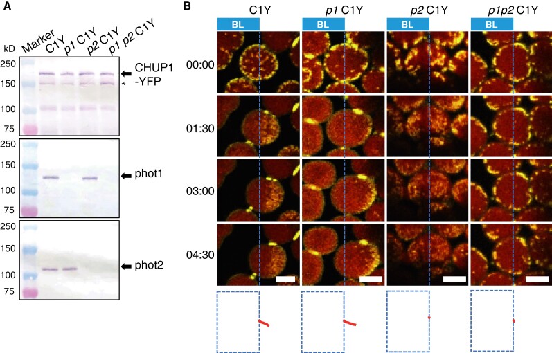
Figure 2.
Identification of the main photoreceptor regulating CHUP1 localization. A) Immunoblot analysis of CHUP1-YFP abundance in transgenic plants in the WT, phot1, phot2, and phot1 phot2 mutant backgrounds (C1Y, p1 C1Y, p2 C1Y, p1 p2 C1Y). Protein samples (50 μg) were separated on 7.5% SDS-PAGE gels and probed with anti-CHUP1 (top panel), anti-PHOT1 (middle), and anti-PHOT2 (bottom) polyclonal antibodies. Arrows from top to bottom blots indicate CHUP1-YFP, phot1, and phot2, respectively. The band indicated by * most likely represents truncated CHUP1-YFP generated by endogenous proteases, since this band is absent in cells lacking CHUP1-YFP. B) Series of images at the indicated time points (min:s) showing the reorganization of CHUP1-YFP in strong blue light in WT and phot mutant cells. In each image, the region to the left of the blue dotted line was irradiated with strong blue light by 458-nm laser scans with an output power of 2.8 μW at 30-s intervals to induce the avoidance response. The images were captured at a resolution of 512 × 256 pixels using a 4× digital zoom and are presented with false-color indicating YFP (yellow) and chlorophyll (red) fluorescence. The time (min:s) of image acquisition is shown to the left. The paths of individual chloroplasts are indicated in red at the bottom of each image. The centers of each chloroplast were traced. Time-lapse movies of these responses can be seen in Supplementary Movies S3 to S6. Scale bars, 5 μm.

