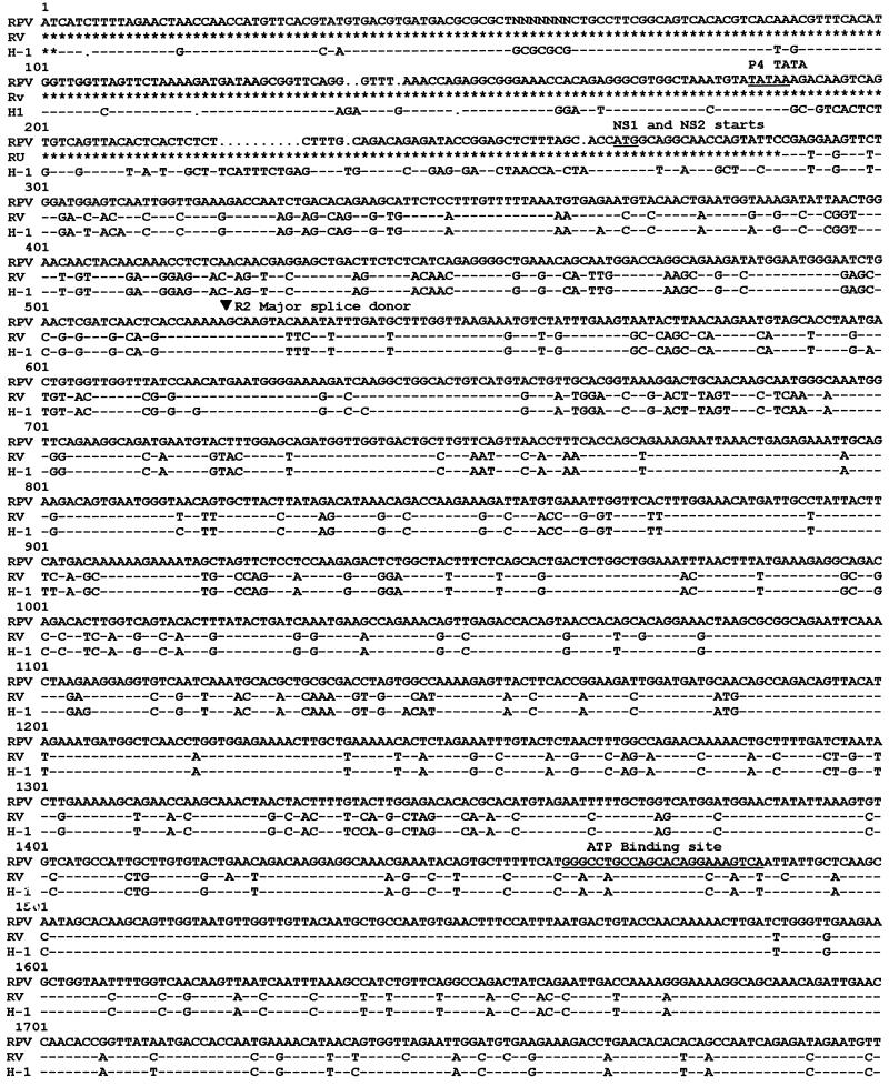
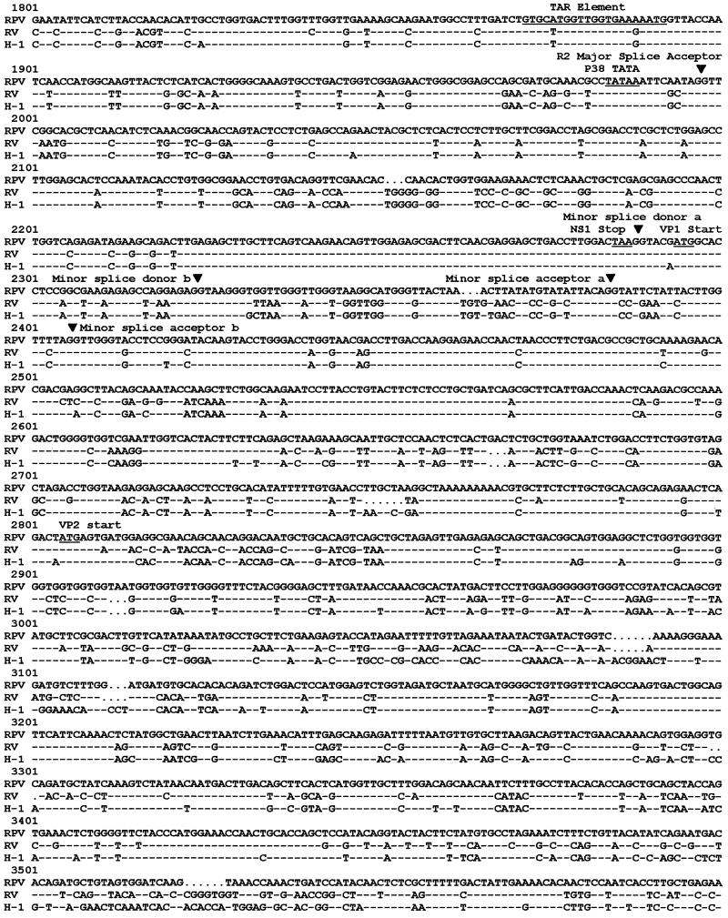
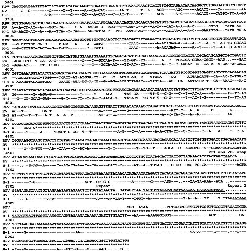
FIG. 1.
Sequence comparison of RPV-1a, RV-UMass, and H-1. The right terminus of H-1 virus is not included, as this region was not cloned or sequenced for RPV-1a or RV-UMass. Dashes indicate nucleotides identical to those of RPV-1a, dots indicate spaces inserted for maximal sequence alignment, and asterisks indicate nonsequenced regions. Regions conserved among parvoviruses are underlined, and MVM splice donors and acceptors are indicated with arrows. GCG programs Pileup and Pretty were used to generate the alignment.


