FIG. 1.
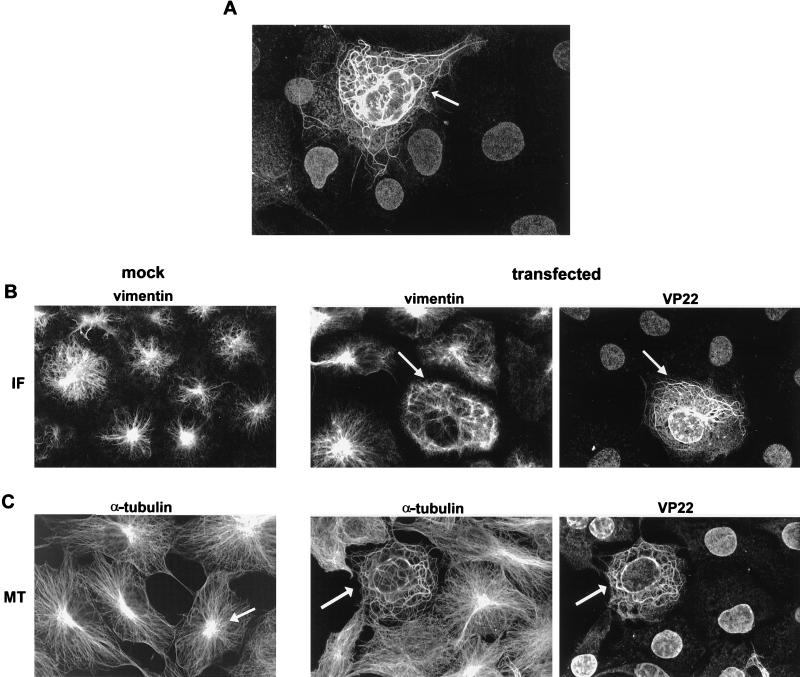
VP22 colocalizes with and reorganizes MTs in transfected cells. (A) Typical VP22 localization pattern in COS-1 cells transfected with plasmid pGE109 and stained with the anti-VP22 polyclonal antibody AGV30. The cell expressing VP22 (arrowed) is surrounded by cells which have taken up VP22 into their nuclei. (B and C) Localization of the cytoskeletal proteins vimentin (B) and α-tubulin (C) in untransfected (mock) and VP22-expressing (transfected) COS-1 cells. Immunofluorescence was carried out with the antivimentin (B) or anti-α-tubulin (C) monoclonal antibodies, which were used in conjunction with AGV30 for double labeling of transfected cells. VP22-expressing cells are arrowed. The cell MT-organizing center is arrowed in panel C, mock.

