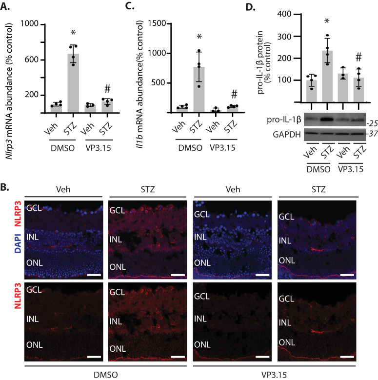
Figure 7.
GSK3 inhibition prevented diabetes-induced NLRP3 inflammasome priming in the retina. Diabetes was induced in mice by administration of STZ. All analyses were performed 16 weeks after mice were administered STZ or a vehicle (Veh). During the last 3 weeks of diabetes, mice were treated daily by intraperitoneal administration of the GSK3 inhibitor VP3.15 or a vehicle (10% DMSO). (A) Nlrp3 mRNA expression was determined in retinal tissue homogenates by RT-PCR. (B) NLRP3 protein (red) was visualized in retinal sections by immunofluorescence. DAPI (blue) was used to visualize nuclei. Representative micrographs are shown (scale bar, 50 µm). (C) Il1b mRNA expression was determined in retinal tissue homogenates by RT-PCR. (D) Pro–IL-1β protein abundance was determined in retinal tissue homogenates by western blotting. Representative blots are shown. Molecular mass in kDa is indicated at right of each blot. Values are means ± SD (n = 3–4). *p < 0.05 vs. Veh; # p < 0.05 vs. DMSO. GCL, ganglion cell layer; INL, inner nuclear layer; ONL, outer nuclear layer.

