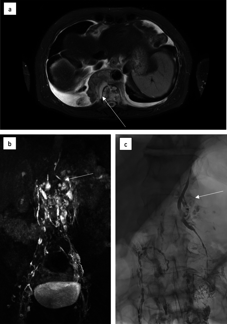
Fig. 1.
MR lymphangiography of a 56-year-old patient with extensive retrocural lymphoma manifestation (white arrow) (a). After nodal contrast application KM ascension via enhancement of pelvic and retroperitoneal lymphatic vessels is visible; in the upper abdomen the lower part of the thoracic duct can be seen. There is no further enhancement of the thoracic duct above the lymphoma mass corresponding to lymphatic obstruction (white arrow) (b). X-ray lymphangiography corroborated obstructive lymphatic drainage disorder at the level of the retrocrural lymphatic mass (white arrow) and consecutive chylolymphatic reflux in the upper abdomen (c)

