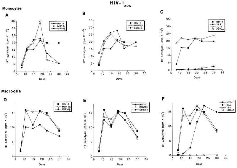
FIG. 5.
Effect of β-chemokine peptides and receptor antibodies on replication profiles of HIV-1ADA. Adherent monolayers of microglia (5 × 10⁴ cells/well in a 96-well plate) and monocytes (105 cells/well) were cultured for 7 days before infection with cell-free stocks of HIV-1ADA. Prior to infection, cells were incubated with antibodies to CCR3 (7B11; 20 μg/ml), CCR5 (3A9; 100 μg/ml), CD4 (OKT4A; 10 μg/ml), or the β-chemokine peptides MIP-1α, MIP-1β, RANTES, and eotaxin (500 ng/ml each) for 1 h at 37°C. Control infected and uninfected cells were maintained simultaneously. The infection proceeded for 4 h after which the virus was washed off and the cells were maintained with media supplemented with appropriate concentrations of antibodies as described in Materials and Methods. Culture supernatant samples were collected twice weekly over a period of 4 weeks postinfection. Each experimental condition was assayed in triplicate, and RT activity was measured independently for each obtained sample.

