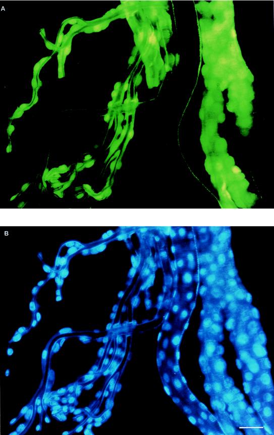
FIG. 2.
(A) Tracheae collected 60 hpi from a T. ni larva infected with AcMNPV-GFP and observed under an Olympus BX50 fluorescence microscope with a fluoresein cube. (B) Same sample as that shown above but stained with DAPI and observed with an Olympus BX50 fluorescence microscope utilizing a DAPI cube. The 1-cm bar in panel B represents 48 μm for both panels.

