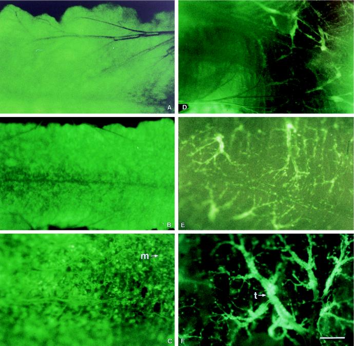
FIG. 3.
Time course of fluorescence within the midguts of AcMNPV-GFP-infected T. ni larvae. Midguts were dissected at 12 (A), 24 (B), 36 (C), 48 (D), 60 (E), and 72 (F) hpi and photographed with a Leica MZ12 dissecting microscope with a fluorescence module and a Leica MPS60 camera with Kodak Ektachrome 160T slide film. The 1-cm bar in panel F represents 100 μm for panels A, B, and C and 88 μm for panels D, E, and F. A representative midgut epithelial cell (m) and tracheae (t) are indicated.

