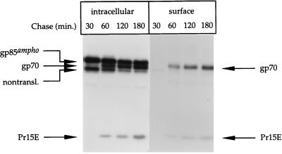
FIG. 2.
Cell surface expression of Env molecules. Amphotropic Mo-MLV env-expressing cells were pulse-labeled and chased as described in the legend to Fig. 1. After the chase, the cells were put on ice and incubated for 30 min with NHS-S-S-biotin. The biotin was then inactivated and washed away. The cells were solubilized in lysis buffer, and the biotinylated proteins (surface) were collected with streptavidin-agarose. The remaining material (intracellular) was subjected to immunoprecipitation with the polyclonal anti-MLV serum. The precipitates were reduced with 50 mM DTT before being subjected to electrophoresis in SDS–12% polyacrylamide gels.

