
Figure 8.
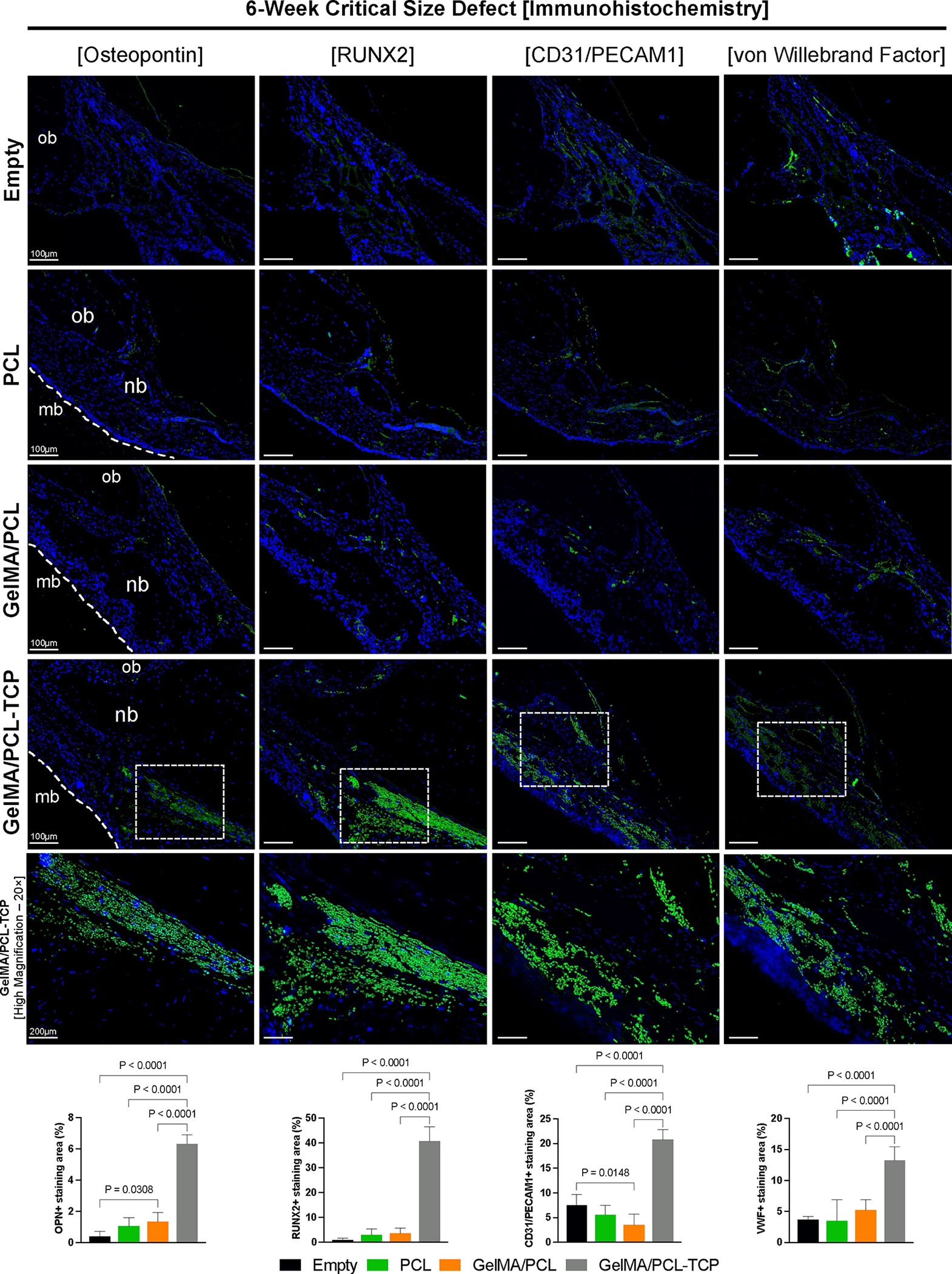
In vivo 6-week rat critical-size defect immunohistochemistry evaluation of bone formation (osteopontin and Runt-related transcription factor 2RUNX2) and angiogenesis (cluster of differentiation 31C—D31 or platelet and endothelial cell adhesion molecule 1—PECAM1 and von Willebrand Factor). For the osteogenic markers, higher immunolabeling is seen for the GelMA/PCL-TCP group, compared to the other three. Even for the empty defect, it is possible to see adequate immunoexpression for angiogenesis-wise markers, similar to the PCL and GelMA/PCL groups; however, undoubtedly, higher immunolabeling can be visualized in the GelMA/PCL-TCP group. Cell nuclei were labeled with DAPI (blue), and antibody binding was visualized using Alexa Fluor 488 (green) secondary antibody. mb: membrane; ob: original bone; nb: new bone. Scale bars: 100 μm and 200 μm for GelMA/PCL-TCP [high magnification]. ImageJ software was utilized to quantify the positively stained area of the four immunomarkers, and they were analyzed by an ordinary one-way ANOVA with Tukey’s multiple comparison test. Bar graphs that exhibited the mean values and their corresponding standard deviations were used to present the results.