
FIGURE 4.
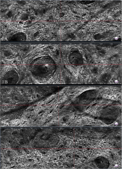
Confocal microscopy, enface, view of LC‐OCT. (1.A) Pre‐HF. (1.B) 10 mins post‐HF (moderate HF, dermis appears stretched as demonstrated by an increase in the size and spacing between collagen fibers). (2.A) Pre‐HF. (2.B) 2 weeks post‐HF (moderate HF, dermis returns to baseline level, there is no visible difference seen in the quantity and quality of collagen fibers as compared to baseline images).