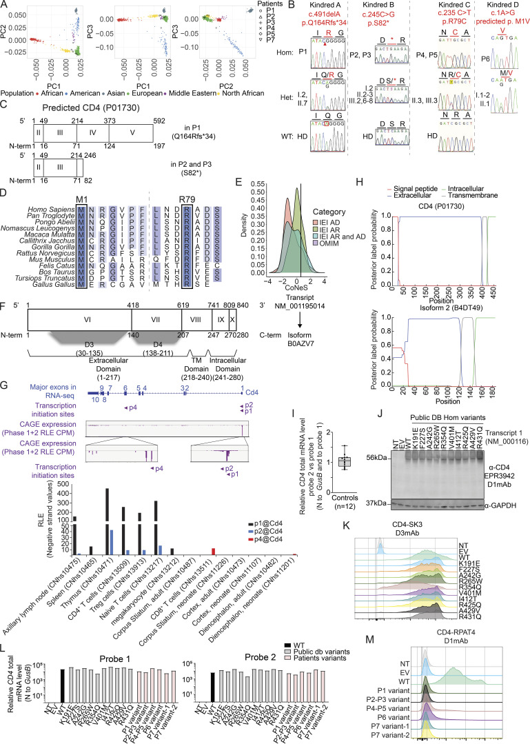
Figure S1.
Genetics, in silico analysis, and impact of identified CD4 variants on mRNA and protein expression. (A) Principal component analysis of WES data from the patients and our in-house WES database. (B) Electropherograms of representative CD4 nucleotide sequences in Kindreds A–D. (C) Schematic representation of the predicted truncated CD4 in P1–P3. (D) Alignment of CD4 M1 and R79 residues in humans and 11 other representative animal species. Dark blue: highly conserved; blue: well conserved; light blue: moderately conserved; white: not conserved. (E) CoNeS of CD4. AD: autosomal dominant, AR: autosomal recessive. (F) Schematic representation of CD4 NM_001195014 and its corresponding isoform (B0AZV7). Exon numeration is based on NM_000616. Nucleotide (above) and amino acid (below) numeration is indicated. Protein domains are represented below isoform. (G) Cd4 transcript expression in mouse tissues. Top: CAGE-seq relative expression track from the FANTOM5 project showing signal pool for all tested tissues. Three transcriptional initiation sites are detected. p1 and p2 are located upstream of exon 1, while p4 is upstream of exon 6. Bottom: Bar graph showing CAGE-seq relative expression for representative tissues. Note that p4 is only detected in the brain. (H) Prediction of transmembrane topologies and signal peptides done by Phobius (https://phobius.sbc.su.se/) based on CD4 and isoform 2 amino acid sequences. Red: signal peptide; blue: extracellular domain; green: Intracellular domain; gray: transmembrane domain. Y axis represents probability, and x axis represents amino acid prediction. (I) CD4 total mRNA level in healthy donors relative to GUS (dCT). For each sample, ddCT was calculated as follows: probe 2 dCT normalized to probe 1 dCT value. (J) Immunoblotting with N-terminal CD4 D1mAb (EPR3942) and GAPDH on total cell lysate from HEK293T either non-transfected (NT) or transiently transfected with an empty vector (EV) or with vectors encoding the indicated CD4 transcript. (K) Flow cytometry following extracellular staining with CD4 (D3mAb; SK3) of HEK293T either non-transfected (NT) or transiently transfected with an empty vector (EV) or with vectors encoding the indicated CD4 transcript. (L) Relative CD4 total mRNA level (probe 1 and 2) normalized to GUS of HEK293T either non-transfected (NT) or transiently transfected with an empty vector (EV) or with vectors encoding the indicated CD4 transcript. (M) Flow cytometry following extracellular staining with CD4 (D1mAb; RPAT4) of HEK293T either non transfected (NT) or transiently transfected with an empty vector (EV) or with vectors encoding the indicated CD4 transcript. Data are representative of at least two independent experiments. Source data are available for this figure: SourceData FS1.

