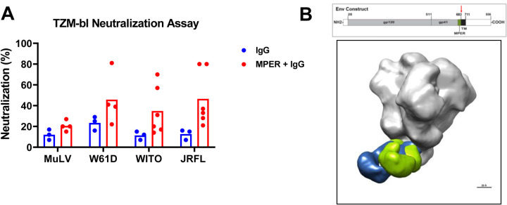
Figure 8. Correlation of Serum for MPER+ NAbs.
(A) Total IgG, and affinity-purified MPER+ IgG, from serum of vaccinee 133–23 and 133–39 post-3rd immunization were tested for neutralization of heterologous tier 1 or tier 2 HIV-1 strains in the TZM-bl assay. MuLV was used as the negative control virus. Each dot represents data from a separate experiment. The bar represents the average of experiments from both vaccinees. Neutralization was reported as percent inhibition at the highest concentration tested. The highest IgG concentration was equilibrated with serum Ig concentration. MPER+ IgG was ~10X serum concentration. P-value=0.024 for JRFL and 0.048 for WITO; Exact Wilcoxon Test. (B) Electron polyclonal epitope mapping (EMPEM) of serum Fab fragments of whole serum IgG. IgG was purified from serum of vaccinee 133–23 after the third immunization, and IgG Fab fragments produced. Negative stained electron microscopy was performed on the Fab-HIV-1 Env trimer complex and demonstrates two Fab fragments overlaid on the trimer, showing binding to the MPER gp41 region. Top diagram illustrates the Env sequence and domain structure. EMPEM was performed with a soluble SOSIP construct that was truncated at residue 683 (red arrow), in between the MPER and the transmembrane domain (TM).

