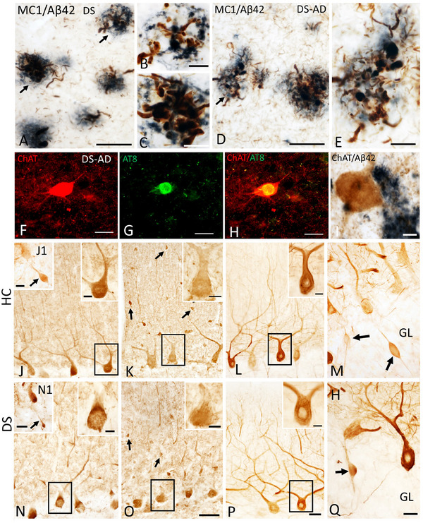
FIGURE 2.

Photomicrographs of dual labeled frontal cortex sections showing dystrophic neurites displaying immunoreactivity for tau conformational epitope MC1 (brown) intermingled within Aβ42‐ir plaques (blue) in a 47‐year‐old female nondemented (A) and a 46‐year‐old male demented (D) individual with DS. Note the presence of numerous MC1 immunoreactive (‐ir) neuropile threads in demented compared to nondemented DS case. (B, C, and E) High‐power images showing bulbous nature of dystrophic neurites within Aβ42‐ir plaques from panels A and D (arrows), respectively. (F–H) Single immunofluorescence images showing normal appearing striatal choline acetyltransferase (ChAT) positive neuron (red, F), AT8‐positive NFT (green; G) in a 46‐year‐old male demented case. (H) Merged image of ChAT and AT8 immunostaining shown in F and G. Note the intact appearance of the cholinergic striatal neuron (red) despite the presence of an AT8 reactivity (yellow) within the perikarya in this demented DS case. (I) Intact ChAT‐positive putaminal neuron (brown) despite its proximity to Aβ42 staining (blue‐black) in a 46‐year‐old male donor with DS and dementia. (J, N) Photomicrographs showing Calb‐ir Purkinje cells (PCs) in a female 66‐year‐old healthy control (HC) (J) and a female 47‐year‐old nondemented DS (N) case. Upper right insets show high‐power image of black‐boxed Calb‐ir PCs in panels J and N. Insets J1 and N1 show cerebellar granular layer (GL) Calb‐ir axonal torpedoes (arrows) in a male 51‐year‐old healthy control (HC) and a female nondemented 60‐year‐old DS case. (K, O) Images showing Parv‐ir PCs and Parv‐ir interneurons (black arrows) within the cerebellar molecular layer (ML) in a female 69‐year‐old HC (K) and a female 44‐year‐old DS without dementia (O). Upper right insets (K, O) are higher‐magnification images of the Parv‐ir PCs shown in the black boxes. (L, P). Photomicrographs of nonphosphorylated high‐molecular‐weight neurofilaments (SMI‐32‐ir) PC dendritic arbors and axons in a female 69‐year‐old HC (L) and a male 46‐year‐old DS‐AD (P) case. Insets in L and P show high‐power images of boxed SMI‐32‐ir PCs and proximal dendrites. (M, Q) Swollen SMI‐32‐ir proximal PC axons or torpedoes (arrows) in GL of male 51‐year‐old HC (M) and female 60‐year‐old DS (Q). Scale bars: A, D, F–H = 50 μm; B, C, E, I and insets in J, K, L, N, O, P = 10 μm; J1 and N1 insets = 30 μm; O = 50 μm and applies to J, K, M; P = 50 μm and applies to L; Q = 25 μm and applies to M.
