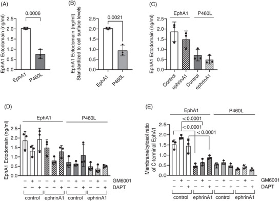
FIGURE 5.

Role of proteolysis in membrane localization and release of soluble receptor by EphA1 and P460L HEK cells under basal and activating conditions. The levels of soluble EphA1 released from unactivated and activated EphA1‐ and P460L‐expressing HEK cells were determined using a human EphA1 ELISA kit that detected the N‐terminal ectodomain (ECD). (A) Total EphA1 ECD released under basal conditions by EphA1 and P460L HEK cells (p = 0.006). (B) Normalized EphA1 ECD released under basal conditions by EphA1 and P460L HEK cells (p =0 .0021). (C) EphA1 ECD released from EphA1‐ and P460L‐expressing HEK cells following incubation with 2 μg/mL sEphrinA1 (ephrinA1) or human IgG (control) for 2 h. (D) EphA1 ECD released from EphA1‐ and P460L‐expressing HEK cells following incubation with ephrinA1 or control in presence of 25 μM GM6001, 25 μM DAPT, or DMSO equivalent for 2 h. (E) Subcellular localization of receptor in EphA1 and P460L HEK cells following incubation with ephrinA1 or control in presence of 25 μM GM6001, 25 μM DAPT, or DMSO equivalent for 2 h by imaging flow cytometry (p = < .0001). Bar charts show mean and error bars indicate ± SD, n = 3.
